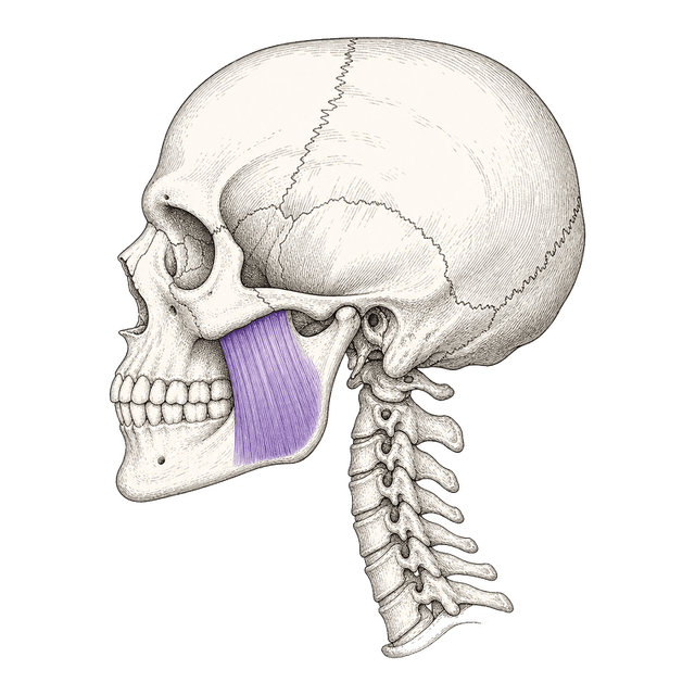
Map the skull. Then reason like the board.
First you label the lateral skull — we name a landmark, you click it. Then eight board-style questions ask what those landmarks do: which muscle attaches, which nerve supplies it, what leaves through each foramen. Options reshuffle every run, and nothing on screen names the answer before you commit.
- ✓Label the lateral skull. Orbit and maxilla, the zygomatic arch, the TMJ landmarks — condyle, coronoid, notch, angle — plus the mental foramen, ear canal, and mastoid. Click roughly where each sits; we score by the nearest structure.
- ✓Then answer the board. Muscles of mastication and their innervation, the TMJ, the mental foramen, the salivary glands, and the cranial nerves of the head and neck — the way the NBDHE asks them.
- ✓Nothing gives it away. A landmark reveals only after you click; question options reshuffle every run and no label names the answer. You commit first, then find out.
- ✓Precision, streak & mastery — and it counts. Land a click dead-on for a Bullseye, build a first-try streak across both halves, and watch every item turn solved. Your score rolls into your Scientific readiness.
10 landmarks + 8 board questions · about 12 minutes
Lateral skull: Gray’s Anatomy (public domain)
Every landmark placed and every board question answered on the first try. Nothing to revisit — this region is locked in.
—
—
This drill is a study aid using a lateral skull plate from Gray's Anatomy (public domain). It builds landmark recognition and tests standard head and neck anatomy — attachments, innervation, and foramina — for self-study, not clinical diagnosis. The mandibular foramen sits on the medial surface of the ramus and is not visible from this lateral view. The House of Hygiene is not affiliated with or endorsed by the JCNDE.